
Postoperative pain is an important consideration in endodontic treatment, particularly in teeth with pulp necrosis and large periapical lesions. A randomized controlled clinical trial published in BDJ Open in 2024 investigated whether an 810 nm diode laser could reduce post-endodontic pain and compared different laser application protocols with conventional treatment.
The findings suggest that combining laser-assisted irrigant activation with low-level laser therapy (LLLT) may help reduce postoperative pain in selected patients. However, the results should be interpreted in the context of the study design, treatment parameters, and limitations.
Key Findings at a Glance
Study design: Four-arm, randomized controlled clinical trial.
Participants: 80 patients with asymptomatic pulp necrosis and large periapical lesions affecting maxillary incisors.
Laser wavelength: 810 nm diode laser.
Interventions: Laser-assisted irrigant activation, low-level laser therapy, combined laser treatment, or conventional treatment without laser application.
Primary outcome: Postoperative pain measured using a visual analog scale (VAS) on days 1, 3, 7, and 14.
Main finding: The combined-treatment group had the lowest mean VAS scores throughout the follow-up period.
Clinical implication: The findings support further investigation of specific 810 nm diode laser protocols as adjuncts to endodontic treatment.
These findings apply to the specific clinical protocol studied and should not be generalized to every diode laser device, wavelength, or endodontic indication.
Study Design and Patient Population
The study, titled Can diode laser 810 nm decrease post endodontic pain in patients with asymptomatic necrotic maxillary incisors? A four-arm randomized controlled trial, was conducted at the Faculty of Dentistry, Damascus University, Syria.
The trial included 80 patients aged 25–44 years. Each patient had a necrotic maxillary central or lateral incisor with an asymptomatic periapical lesion larger than 5 mm. All participants underwent standardized single-visit root canal treatment and were randomly assigned to one of four groups, with 20 patients in each group.
Postoperative pain was evaluated using a 10 cm visual analog scale (VAS), ranging from no pain to unbearable pain. Assessments were performed 1, 3, 7, and 14 days after treatment.
The study protocol was approved by Damascus University and registered in the ISRCTN clinical trial registry under identifier ISRCTN99457940.
The Four Treatment Protocols
Group 1: Conventional Endodontic Treatment Without Laser Application
The control group received standardized root canal treatment without diode laser irradiation. Irrigant activation was performed using an ultrasonic device. This group served as the reference for evaluating the effects of the laser-assisted protocols.
Group 2: Laser-Assisted Irrigant Activation
An 810 nm diode laser was used to activate root canal irrigants. A 200 μm optical fiber was positioned approximately 2 mm short of the apical endpoint.
The laser-assisted protocol was used with sodium hypochlorite, EDTA, and chlorhexidine according to the study protocol.
The reported laser settings are not detailed here because some of the technical parameters in the publication appear inconsistent and require clarification.
Group 3: Low-Level Laser Therapy
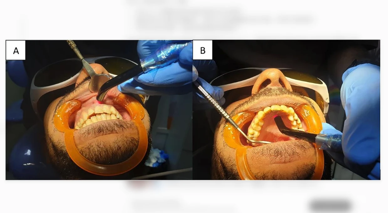
After root canal treatment and restoration, an 810 nm diode laser was applied externally over the apical region through the buccal and palatal mucosa.
The reported parameters were:
Output power: 100 mW
Application distance: 3 mm from the oral mucosa
Application duration: 80 seconds per side
Application sites: Buccal and palatal sides
This protocol investigated the potential effect of low-level laser therapy on postoperative pain.
Group 4: Combined Laser-Assisted Irrigant Activation and Low-Level Laser Therapy
The fourth group received both laser-assisted irrigant activation and the external low-level laser treatment described above. This approach allowed the investigators to compare the combined protocol with either laser technique used independently.
What Did the Researchers Find?
The researchers reported statistically significant differences in mean postoperative VAS scores among the four groups at all four follow-up assessments. The combined-treatment group had the lowest mean pain scores throughout the 14-day follow-up period.
The table below summarizes the mean VAS scores and standard deviations reported in the original publication.
| Assessment | No Laser | Irrigant Activation | LLLT | Combined Treatment |
|---|---|---|---|---|
| Day 1 | 5.00 ± 2.05 | 3.20 ± 1.61 | 0.80 ± 0.63 | 0.20 ± 0.42 |
| Day 3 | 2.80 ± 1.68 | 1.60 ± 0.96 | 0.40 ± 0.51 | 0.00 ± 0.00 |
| Day 7 | 0.80 ± 0.91 | 0.40 ± 0.69 | 0.00 ± 0.00 | 0.00 ± 0.00 |
| Day 14 | 0.40 ± 0.51 | 0.20 ± 0.42 | 0.00 ± 0.00 | 0.00 ± 0.00 |
Data source: Abbara et al., BDJ Open, 2024, Table 3. Values are reported as mean ± standard deviation.
Day 1: The Largest Differences Were Observed
On the first day after treatment, the mean VAS score was 5.00 in the control group, compared with 3.20 in the laser-assisted irrigant activation group, 0.80 in the LLLT group, and 0.20 in the combined-treatment group.
The authors reported statistically significant pairwise differences among all four groups at this assessment.
Day 3: The Combined Group Maintained the Lowest Score
On day 3, the combined-treatment group had a mean VAS score of 0.00. The LLLT group had a mean score of 0.40, the irrigant activation group 1.60, and the control group 2.80.
However, the difference between the control and irrigant activation groups was not statistically significant at this time point (P = 0.072).
Days 7 and 14: Pain Scores Were Low Across the Laser Groups
By day 7, mean VAS scores had decreased to 0.40 in the irrigant activation group and 0.00 in both the LLLT and combined-treatment groups. On day 14, the corresponding mean scores were 0.20, 0.00, and 0.00.
The pairwise analyses found no statistically significant differences among the three laser groups on days 7 and 14. The LLLT and combined-treatment groups had significantly lower scores than the control group at both assessments.
Statistical Significance and Clinical Interpretation
The overall between-group comparisons were statistically significant on day 1 (P < 0.001), day 3 (P < 0.001), day 7 (P = 0.004), and day 14 (P = 0.038).
The results indicate that the combined protocol produced the lowest observed mean pain scores. Nevertheless, a lower score in this trial does not establish that the same benefit will occur in all patients or with other laser systems.
VAS is a subjective outcome measure. The study did not establish whether the observed differences in postoperative pain translate into improved long-term healing of periapical lesions.
How Might Diode Laser Therapy Influence Postoperative Pain?
The researchers discussed two potential mechanisms that may help explain the observed findings.
1. Laser-Assisted Irrigant Activation
During root canal treatment, irrigants help clean the canal system and reduce microbial contamination. Laser irradiation can generate fluid movement and promote irrigant agitation under appropriate operating conditions.
The authors proposed that improved irrigant activity could contribute to more effective cleaning and reduced irritation of periapical tissues. This provides a plausible explanation for the lower pain scores observed in the laser-assisted irrigant activation group.
However, the trial primarily evaluated postoperative pain. It did not directly establish that changes in microbial load or irrigant penetration were responsible for the differences in pain scores.
2. Low-Level Laser Therapy and Photobiomodulation
Low-level laser therapy, also known as photobiomodulation (PBM), uses light at controlled parameters to interact with biological tissues.
Potential mechanisms discussed in the literature include modulation of inflammatory responses, changes in cellular metabolism, and effects on pain-related signaling pathways.
In this study, the laser was applied externally over the apical region rather than inside the root canal. The investigators proposed that this approach may help modulate the local tissue response following endodontic treatment.
These mechanisms are biologically plausible, but they were not directly measured in this trial. Further research is needed to determine which mechanisms account for the observed clinical outcomes.
Limitations of the Study
Although the findings are promising, several limitations should be considered before applying them to routine clinical practice.
Limited sample size: The study included 80 patients, with 20 participants in each group. Larger, independently replicated trials would help establish the reliability and generalizability of the findings.
Narrowly defined patient population: The study involved asymptomatic necrotic maxillary incisors with large periapical lesions in patients aged 25–44 years. The results may not apply to symptomatic teeth, molars, other lesion sizes, or different clinical conditions.
Limited blinding: The trial was single-blinded. The treating clinician could not be blinded to the intervention, and patients receiving external laser irradiation were informed about the procedure. Although outcome assessors were blinded to the laser application method, awareness of treatment could still influence subjective pain reporting.
Short follow-up period: Pain was monitored for 14 days. The trial did not establish the effects of these protocols on long-term periapical healing, tooth survival, or other long-term clinical outcomes.
Subjective primary outcome: Pain was assessed using the VAS. Although widely used in clinical research, this measure reflects patients' perceptions and can be influenced by factors other than the intervention.
Protocol-specific findings: The study used defined laser settings, delivery methods, and irrigant protocols. Its results should not be assumed to apply to devices with different wavelengths, power settings, fiber dimensions, or application techniques.
Further studies involving placebo-controlled designs, double blinding where feasible, and different pulp and periapical conditions are needed. Additional research is also required to investigate whether laser application methods influence periapical lesion healing.
What This Study Means for Dental Laser Practitioners
From a clinical perspective, this trial provides evidence that the application method may be an important consideration when evaluating diode laser use for postoperative pain management.
The study suggests three practical areas for further investigation:
Irrigant activation: Assessing whether laser-assisted activation offers benefits under specific endodontic protocols.
Photobiomodulation: Investigating the potential contribution of external low-level laser therapy to postoperative comfort.
Combined protocols: Evaluating whether integrating intracanal and external laser applications offers clinically meaningful advantages over either approach alone.
The findings should be viewed as evidence for a particular treatment protocol, not as a universal recommendation. Treatment decisions should consider the clinical diagnosis, the patient's needs, the device instructions for use, and the clinician's training.
Frequently Asked Questions
Can an 810 nm diode laser reduce pain after root canal treatment?
In the randomized controlled trial discussed here, laser-assisted irrigant activation, low-level laser therapy, and their combination were associated with lower postoperative VAS scores than the no-laser control group at selected follow-up assessments. The results are encouraging but require confirmation in further studies.
Which laser application method produced the lowest pain scores?
The combined protocol, consisting of laser-assisted irrigant activation and external low-level laser therapy, had the lowest mean VAS scores in this trial. However, the study did not demonstrate statistically significant differences among the three laser groups on days 7 and 14.
Is 810 nm the best wavelength for postoperative pain management?
This study evaluated one wavelength, 810 nm. It did not compare multiple wavelengths, so it cannot establish that 810 nm is superior to other wavelengths for postoperative pain management.
Does the study prove that diode lasers improve periapical healing?
No. The primary outcome was postoperative pain, assessed over 14 days. Long-term periapical healing was not established by this trial.
Can the findings be applied to every dental diode laser?
Not directly. Clinical outcomes depend on the wavelength, output parameters, delivery system, treatment technique, patient population, and clinical indication. Practitioners should follow the applicable device instructions and use evidence relevant to the intended application.
Conclusion
This four-arm randomized controlled trial provides clinical evidence that specific 810 nm diode laser protocols may help reduce postoperative pain following single-visit root canal treatment in patients with asymptomatic necrotic maxillary incisors and large periapical lesions.
The combined protocol produced the lowest mean VAS scores throughout the 14-day follow-up. Nevertheless, the limited sample size, single-blinded design, narrowly defined patient population, and short follow-up period mean that the findings should be interpreted cautiously.
Further research is needed to determine whether these results can be reproduced in broader patient populations and whether the observed reductions in postoperative pain are associated with improvements in long-term endodontic outcomes.
Original Research and References
Abbara MT, Akil S, Hamadah O, Achour H, Mahayni G, Alsayed Tolibah Y. Can diode laser 810 nm decrease post endodontic pain in patients with asymptomatic necrotic maxillary incisors? A four-arm randomized controlled trial. BDJ Open. 2024;10:23.
DOI: https://doi.org/10.1038/s41405-024-00203-w
Full text: Read the full study on PubMed Central
PubMed: View the PubMed record
Source attribution: The clinical findings and experimental parameters presented above are based on the cited original research. The clinical interpretation and practitioner-oriented discussion are editorial analysis and should not be interpreted as additional experimental findings.
Device disclosure: The original study reports using the Mercury G10 810 nm diode laser manufactured by Wuhan Pioon Technology Co., Ltd. This identifies the device used in the study; it does not establish that every PIOON laser model has been evaluated for the same indication or that the study's results can be generalized to other devices.
Medical information notice: This article is an educational interpretation of published research and is not a substitute for professional clinical judgment, patient-specific diagnosis, or the manufacturer's instructions for use.