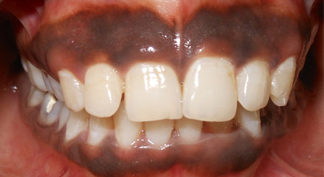
Introduction - Gingival hyperpigmentation is a common esthetical concern in patients with gummy smile or excessive gingival display. It is an overproduction of melanin, beyond the normal expected degree in the oral mucosa, induced by various causes.
Etiology - Several physiologic and/or pathologic factors can cause hyperpigmentation. Gingival hyperpigmentation is mostly caused by the physiologic deposition of a brown pigment, melanin produced by melanocytes in the basal and supra-basal cell layer of the gingival epithelium. The colour of gingiva depends on the intensity of the melanogenesis, degree of epithelial cornification, depth of epithelialization and the arrangement of gingival vascularity.
Treatment Modalities – Gingival depigmentation is a treatment to remove melanin hyperpigmentation of the gingiva and various methods have been used for this procedure with different degrees of success including gingivectomy, gingivectomy with free gingival autografting, electrosurgery, cryosurgery, chemotherapy with 90% phenol and 95% alcohol and abrasion with diamond bur. Moreover some of these techniques are prone to side effects and complications.
However, Laser therapy is an effective and noninvasive treatment option for Gingival Depigmentation. In depigmentation process the epithelial tissue exhibiting excessive melanin pigments is ablated using diode lasers. PIOON Laser offers different wavelengths like 450nm, 810nm/980nm which can either be used in contact or non-contact manner to perform this procedure.
Most recommended for an effective treatment procedure is the blue light (450nm) which can be backed by a report by Kenneth Luk in 2017 where he studied the comparison between the 810nm and 445nm working in a noncontact mode to perform depigmentation of gingiva. He stated that 445nm is much better absorbed by melanin and haemoglobin than 810 nm and hence, a much lower power density can be used for the treatment. Manaf Agha in 2020 concluded that lesser the power settings used for the procedure, faster would be the healing with much less discomfort to the patient leading to greater acceptance of the procedure.
Before laser irradiation, the operating staff, assistant and the patient wears special laser-protective eye glasses corresponding to laser wavelength, highly reflective instruments or instruments with mirrored surfaces are avoided and most of the times only the use of topical anesthesia suffices for a painless laser tissue ablation.
Rationale behind Use of Lasers - Laser ablation has been recognized as a pleasant and reliable technique. It has the advantage of easy handling of tissues because of bloodless field, shorter chairside time, treatment finishes in single visit and property of potential decontamination. Additionally, protein coagulum formed on the depigmented surface as a result of irradiation act as a biological wound dressing sealing the ends of sensory nerve endings and hence lesser pain and discomfort is experienced by the patients.
Conclusion - Diode dental laser is a safe and effective treatment modality that provides optimal aesthetics with minimal discomfort to patients with gingival hyperpigmentation.
Reference – Luk K. Non-ablative melanin depigmentation of gingiva. Cosmetic dentistry. 2017. 36-39 and Agha M. Laser Treatment for Melanin Gingival Pigmentations: A Comparison Study for 3 Laser Wavelengths 2780, 940, and 445nm. Int J Dent.2020. March: 3896386.

By – Dr. Sana Farista
MDS Periodontics & Laser Specialist