Introduction: Understanding Periodontal Disease
A periodontal disease starts when the bacteria, (often found in plaque), infect the areas around the teeth and form the hardened substance called calculus. This leads to the inflammation of gingiva and affects both the tooth and underlying jawbone (called as Periodontitis).
Challenges in Conventional Treatment
The initial therapy involves periodontal curettage/debridement to remove plaque and calculus. However, this method doesn't eliminate bacteria effectively, resulting in chronic recurrence. As per the periodontal consensus, conventional periodontal curettage is no longer advised.
Laser Technology as a Revolutionary Solution
To address this challenge, laser technology proves to be a game-changer. Laser Curettage/Debridement effectively eliminates bacterial colonies thriving in pockets at the base of teeth, promoting healing of diseased gums and teeth.
Surgical Procedure in Laser Curettage/Debridement
The laser procedure consists of three key steps:
Pocket Depth Measurement: Using a periodontal probe to measure the depth of pockets.
Scaling and Root Planing: The removal of calculus and root planing for optimal oral health.
Laser Application: Utilizing a laser to perform Curettage/Debridement, targeting bacterial colonies.
Detailed Procedure of Laser Curettage/Debridement
In the presented case of chronic periodontitis:
Topical Anesthesia: Before the laser procedure, topical anesthesia is applied for patient comfort.
Laser Application: PIOON S3 Laser with a 980nm wavelength is used at a power setting of 1-Watt in continuous wave mode. The 300-micron tip is carefully inserted into the pocket, removing the pocket lining epithelium in a continuous movement within a specified time frame.
Scientific Perspectives on Laser Curettage
An overview by Douglas N in 2002 provided insights into the scientific consensus on laser curettage, discussing its effectiveness and potential for collateral damage. Research by Elavarasu S et al in 2015 highlighted the enhanced reduction in Periodontal Depth and Clinical Attachment Level when laser curettage was used as an adjunct to SRP (Scaling and Root Planing).
Conclusion: Embracing Diode Lasers for Effective Treatment
Diode lasers have emerged as promising tools in modern minimally invasive dentistry, demonstrating success in treating periodontal pockets. Their effectiveness in conjunction with scientific standards positions them as a valuable asset in the evolving landscape of periodontal care.
REFERENCE - Douglas N Dederich. Laser curettage: an overview. Compend Contin Educ Dent 2002 Nov;23(11A):1097-103 and Elavarasu S et al. LASER curettage as adjunct to SRP, compared to SRP alone, in patients with periodontitis and controlled type 2 diabetes mellitus: A comparative clinical study. J Pharm Bioallied Sci. 2015 Aug; 7(Suppl 2): S636–S642.

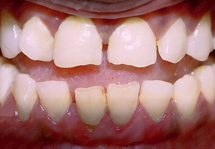
Preoperative View with Red Inflammed Lower Gingiva (Case Courtesy – Dr. Sana Farista)

Postoperative View with Non-Inflammed Lower Gingiva (Case Courtesy – Dr. Sana Farista)
- Dr Aditi Chaudhary
MDS Periodontist
Divas in Laser